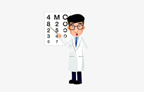
A regular eye examination is an important preventive measure to protect our vision as many of the sight-threatening eye diseases have no obvious symptoms. Only by early diagnosis, early referral and appropriate treatment, our vision can be saved. Comprehensive eye exams can detect diseases and disorders such as glaucoma, cataracts, retinal detachments and macular degeneration, as well as other systemic health problems such as diabetes and high blood pressure.
A comprehensive eye exam includes:
1. History Taking
We will ask about your visual problems, the history of the visual problem such as past investigations and treatment that had been received. We will also ask about your medical, drug and family history to gather information on how these factors are related to your presenting complaint. Besides, an analysis of the patient’s visual needs at home, work, school and play. In some instances, this may necessitate questions about the patient’s school/work environment and recreational activities, in order to accurately determine the patient’s visual demands.
2. Preliminary tests
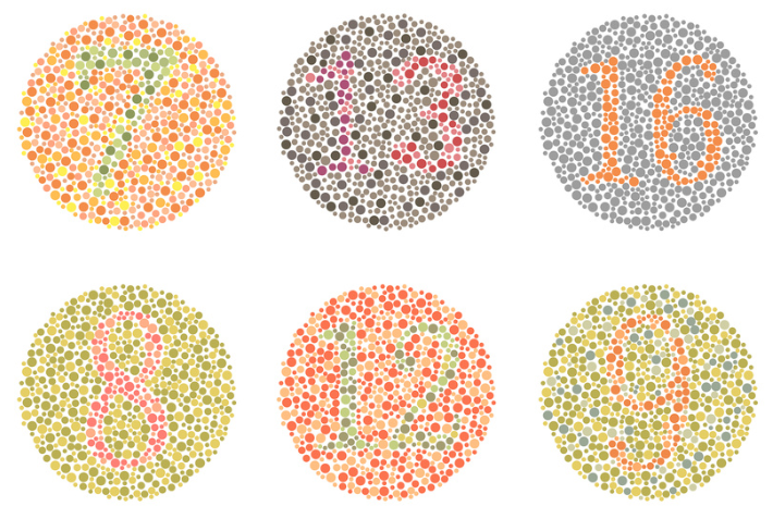
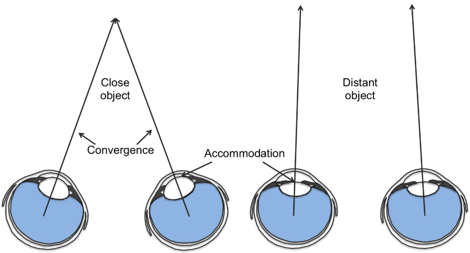
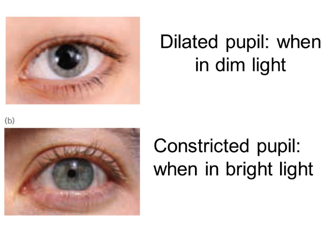
These include evaluation of colour vision, depth perception, eye muscle movement, pupil response and visual acuity. This fundamental information provides us with how good your eyes are functioning and coordinating with each other. Further investigation will then focus on the weaker part of your visual system.
3. Refraction
Refraction or subjective refraction determines the lens power that you need to correct your short-sightedness, long-sightedness, astigmatism and presbyopia. Starting with a quick Auto-refraction, a preliminary refractive status of your eyes is measured. We will then refine the refraction by adding lenses in front of your eyes and base on your responses, the subjective refraction is determined.
We will also use retinoscopy to undergo refraction for younger children. Based on the way the light reflects from your eye, we are able to estimate the power of eyeglasses required to correct your vision. This test is especially useful for children and patients who are unable to accurately answer the optometrist’s questions.
4. Eye Health Evaluation
Our eyeball is basically divided into 2 segments: the anterior segment which corresponds to the front 1/3 of the eye that includes the cornea, iris, ciliary body and the crystalline lens. The posterior segment, the back 2/3 of the eye, includes vitreous humor, retina, choroid and optic nerve.
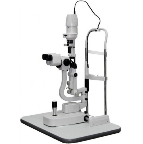
To evaluate the anterior segment, we will use a slit lamp to specifically focus on a particular structure to see if there is abnormal change. Conditions such as dry eyes, corneal opacity and cataract etc. can be investigated using this technique. Slit lamp can also be used in evaluating external eye structures like eye lid and eye lashes.
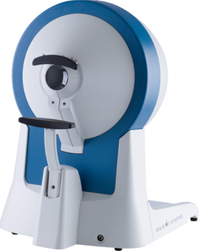
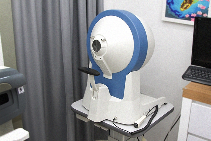
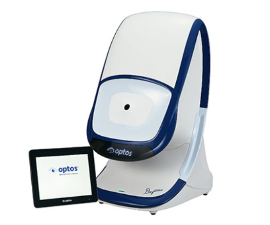
To evaluate the back of the eye, the posterior segment, we will use an instrument –Optos Daytona. The Optos Daytona could allow us to have a 200˚ view of the retina with just a single capture. The Daytona brings the benefits of ultra-widefield optomap images to us for a better view of the retina and the macula. Retinal disease or conditions like age-related macular degeneration (AMD), retinal degeneration (hole, breaks or tears etc.), diabetic retinopathy, retinal vein/artery occlusion, posterior vitreous detachment, floaters and optic nerve atrophy will be investigate.
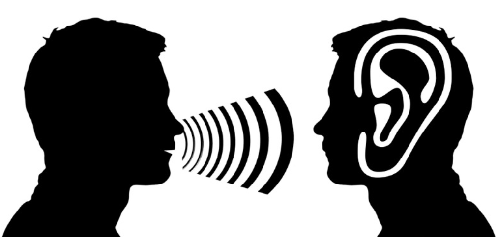
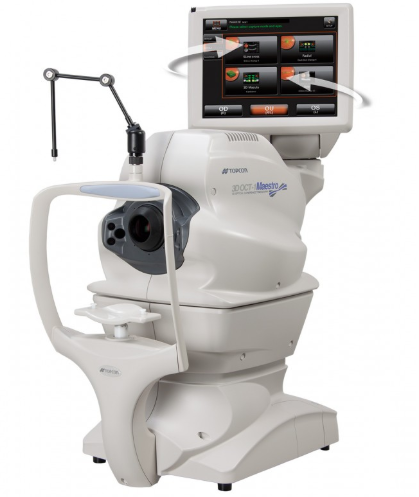
Besides, an Optical Coherence Tomography (OCT) scan will also be done. OCT, similar to ultrasound, this diagnosis technique employs light rather than sound waves to achieve higher resolution pictures of the structural layers of the back of the eye. With an OCT scan, macular hole, macular edema, epiretinal membrane, central serous retinopathy and the anterior chamber angle could be examined. The optic nerve head also can be examine using an OCT. Examination on the optic nerve head also provides us with important information about potential glaucoma, inflammation of the optic nerve or even potential tumour at the back of the eyeball.
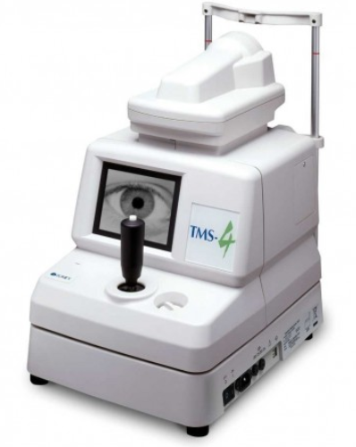
Other than that, a corneal mapping topographer will be used to detect keratoconus and corneal disease. Besides that, it can also be used for contact lens fitting and ortho-keratology fitting that helps in myopia control. Therefore, an evaluation at the back of the eye is an essential part of an eye examination.
5. Eye pressure measurement
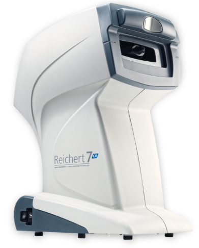
Intraocular pressure (IOP) measurement or tonometry is an evaluation of the pressure inside the eye. An IOP of 10-21mmHg is generally considered as normal. Too high the pressure may cause glaucoma – a disease with progressive loss in the visual field and eventually lead to blindness. Checking the IOP is the first line screening for glaucoma. It can be done easily by a non-invasive method called non-contact air puff test. Our Reichert 7 IOP measurement even able to detect on post-Lasik Patient accurately.
6. Additional tests
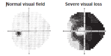
Depends on the individual results, additional test may be needed to further investigate the condition of the eye. For example, a visual field screening test will be suggested if there is family history of glaucoma, or the intraocular pressure (IOP) reading is on the high side. A visual field test is an eye examination that can detect dysfunction in central and peripheral vision which may be caused by various medical conditions such as glaucoma, stroke, pituitary disease, brain tumours or other neurological deficits.